Circle of Willis (CoW) Annotation
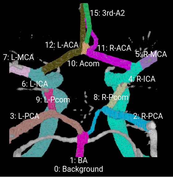
The vessel components of the CoW annotated are left and right internal carotid artery (ICA), left and right anterior cerebral artery (ACA), left and right middle cerebral artery (MCA), anterior communicating artery (Acom), left and right posterior communicating artery (Pcom), left and right posterior cerebral artery (PCA), and basilar artery (BA).
Note that only vessel components and regions necessary to diagnose the CoW angio-architecture and variants are annotated. (See below "CoW ROI 3D Bounding Box" for more info)
Here is the annotation mask in pixel value for each vessel objects of interest for a MRA example:
For the binary segmentation task, the vessel components from above are combined into two classes:
- 0: Background
- 1: CoW Vessels
Annotation & Verification Protocol
Initial CoW annotations are manually labeled voxel-wise by research staff who have gone through CoW anatomy education from the clinical experts. The cases that annotators are uncertain are flagged, and sent for further verification and approval by the clinical experts. The clinical expert team consists of neuro-radiologists, neurologists, and neurosurgeons.
The annotation protocol on how to segment vessel components at bifurcation points such as ACA- ICA-MCA, ACA-Acom, PCA-Pcom, Pcom-ICA, etc., are discussed and agreed upon by the clinical experts. For example, we mark the superior tip of the bifurcations to be part of ICA and BA (due to aneurysm classification). We also include the infundibulum as the origin of the vessel components. The annotation protocol also covers CoW variants such as fetal PCA, triple ACA etc.
Joint-Modalities CTA and MRA
All patients have both CTA and MRA modalities, one scan for each modality. The joint-modality aspect is that for each MRA, there is a corresponding CTA pair from the same patient. Joint-modalities from the same patient will serve as additional reference and provide supplementary anatomical information on the CoW vessels.
Annotators and clinical experts have both CTA and MRA modalities available to annotate and verify. All MRA scans are checked with the paired CTA scan and vice versa to make use of the supplementary source of information from both modalities during annotation. We believe our joint-modality annotations on MRA and CTA can ensure good quality anatomical annotations.
CoW ROI 3D Bounding Box
We also annotate 3D bounding boxes for CoW region of interest (ROI). The CoW ROI is defined as the 3D bounding box capturing the anatomical variant of the CoW. The annotated CoW ROI is released for all training cases. Note that the evaluation of segmentation results is limited to the CoW region of interest.
